Read Heart Mechanics: Magnetic Resonance Imaging—Advanced Techniques, Clinical Applications, and Future Trends: Volume 2 - El-Sayed H. Ibrahim | ePub
Related searches:
Morphological and functional evidences of the helical heart from non
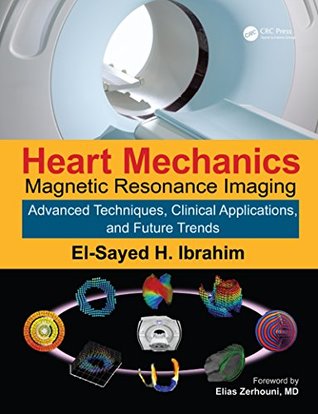
Heart Mechanics: Magnetic Resonance Imaging—Advanced Techniques, Clinical Applications, and Future Trends: Volume 2
Magnetic Resonance Imaging and Ventricle Mechanics - JSTOR
Myocardial Architecture, Mechanics, and Fibrosis in - Frontiers
Heart mechanics and metabolism - The University of Auckland
Magnetic Resonance Imaging (MRI) - MedStar Heart and Vascular
The Magnetic Field Produced by the Heart and Its Influence on MRI
Cardiac Magnetic Resonance Imaging - Brigham and Women's Hospital
(PDF) Magnetic resonance imaging and ventricle mechanics
Advanced echocardiography and cardiac magnetic resonance in
Magnetic Resonance Imaging (MRI) of the Heart - Heart and
During the past decade, cardiovascular magnetic resonance myocardial feature tracking has emerged as a useful tool for the quantitative evaluation of cardiovascular function. The method allows quantification of biatrial and biventricular mechanics from measures of deformation: strain, torsion, and dyssynchrony.
Cardiac magnetic resonance imaging (cardiac mri) produces detailed images of the beating heart.
Patients with prosthetic valves (mechanical or bioprosthetic) or coronary stents may have an indication to undergo magnetic resonance imaging (mri). Sometimes these patients are excluded from mri on the basis that they have an implant that makes them unsuitable for the magnetic resonance (mr) environment.
Cardiovascular imaging is key for the assessment of patients with heart failure. Today, cardiovascular magnetic resonance imaging plays an established role in the assessment of patients with suspected and confirmed heart failure syndromes, in particular identifying aetiology.
Magnetic resonance imaging (mri) is a test that uses a large magnet, radio signals, and a computer to make images of organs and tissue in the body.
Magnetic resonance angiography–also called a magnetic resonance angiogram or mra–is a type of mri that looks specifically at the body’s blood vessels. Unlike a traditional angiogram, which requires inserting a catheter into the body, magnetic resonance angiography is a far less invasive and less painful test.
Heart mechanics by magnetic resonance imaging: techniques and applications� saturday, 16 april, 2:00pm-5:45pm.
Aug 21, 2019 considering the different pathophysiological processes in hhd and hcm and their related cardiac mechanics, we hypothesized that cmr-ft.
Tagged cardiac magnetic resonance imaging (mri) produces images of the heart which can be analyzed to yield detailed maps of motion and mechanical strain, describing regional function of the heart.
Cardiovascular magnetic resonance (cmr) and echocardiography are both commonly used for these studies [ 1, 2 ], however cmr is generally considered to be the more accurate modality. Using cine imaging, left ventricular (lv) volume, stroke volume, ejection fraction, and wall thickening can be quantified.
Cardiac magnetic resonance imaging (cmr) adds valuable additional generalization of quantification of myocardial mechanics either with cmr-ft or other.
Magnetic resonance imaging and ventricle mechanics june 2001 philosophical transactions of the royal society a mathematical physical and engineering sciences 359(1783):1263-1275.
Magnetic resonance imaging (mri) is a medical imaging technique used in radiology to form pictures of the anatomy and the physiological processes of the body. Mri scanners use strong magnetic fields, magnetic field gradients, and radio waves to generate images of the organs in the body.
May 23, 2017 1adult congenital heart unit, royal brompton hospital, london, uk; 2 cardiovascular magnetic resonance unit, royal brompton hospital,.
Question what are the cardiovascular effects in unselected patients with recent coronavirus disease 2019 (covid-19). Findings in this cohort study including 100 patients recently recovered from covid-19 identified from a covid-19 test center, cardiac magnetic resonance imaging revealed cardiac involvement in 78 patients (78%) and ongoing myocardial inflammation in 60 patients (60%.
Heart failure (hf) can be defined haemodynamically as any abnormality of cardiac structure or function resulting in a failure to deliver oxygen at a rate adequate for tissue requirements, despite normal filling pressures – or only at the expense of increased filling pressures. 1 around half of patients with hf have reduced left ventricle ejection fraction (lvef; ef 40 %) at rest (hf-ref).
Recently, cardiovascular magnetic resonance (cmr) has emerged as an important imaging technique, particularly well-suited to provide detailed characterization of the heart and an important aid for diagnosis of underlying heart disease in athletes. 5 cmr provides 3-dimensional tomographic imaging with high spatial and temporal resolution, with the ability to image the heart in any plane and without ionizing radiation.
A test that produces high-quality still and moving pictures of the heart and great vessels. Mri uses large magnets and radio-frequency waves to produce pictures of the body’s internal structures; no x-ray exposure is involved.
With magnetic resonance imaging (mri), a powerful magnetic field and radio waves are used to produce detailed images of the heart and chest. This expensive and sophisticated procedure is used predominantly for the diagnosis of complex heart disorders that are present at birth (congenital) and to differentiate between normal and abnormal tissue.
Mri techniques have been recently introduced for non-invasive qualification of regional myocardial mechanics, which is not achievable with other imaging modalities.
Purpose of test: cardiac magnetic resonance imaging (mri) is a diagnostic procedure that uses a combination of a large magnet, radiofrequencies, and a computer to produce detailed still and moving images of the heart (there is no radiation).
Jun 18, 2015 disparate patterns of left ventricular mechanics differentiate constrictive pericarditis from restrictive cardiomyopathy.
An american heart association scientific statement from the committee on diagnostic and interventional cardiac catheterization, council on clinical cardiology, and the council on cardiovascular radiology and intervention: endorsed by the american college of cardiology foundation, the north american society for cardiac imaging, and the society for cardiovascular magnetic resonance.
Because cardiac magnetic resonance imaging also acquires information about the heart rhythm, it can create clear moving images of the heart throughout its pumping cycle. This allows cardiac magnetic resonance imaging to display abnormalities in cardiac chamber contraction and to show abnormal patterns of blood flow in the heart and great vessels.
Based on research and clinical trials, this book details the latest research in magnetic resonance imaging (mri) tagging technology related to heart mechanics. It covers clinical applications and examines future trends, providing a guide for future uses of mri technology for studying heart mechanics.
We sought to investigate the association between lv mechanics and the presence, location, and extent of fibrosis in patients with nonischemic cardiomyopathy. Methods and results we retrospectively identified 239 patients with nonischemic cardiomyopathy (67% male; 55±14 years) referred for a clinical cardiovascular magnetic resonance.
Effects of magnetic fields used in mri on 15 prosthetic heart valves. Safety of magnetic resonance imaging in patients with cardiovascular.
Tissue tracking technology of routinely acquired cardiovascular magnetic resonance (cmr) cine acquisitions has increased the apparent ease and availability of non-invasive assessments of myocardial deformation in clinical research and practice.
The depiction of myocardial fiber orientation is fundamental to understand ventricular mechanics.
Magnetic resonance imaging, better known as mri, creates detailed pictures of the heart and nearby blood vessels using a combination of radio waves, magnets and computer technology. These pictures help us assess your heart’s structure and function without using a contrast dye or radiation.
Noninvasive imaging plays a central role in the diagnosis of heart failure, assessment of prognosis, and monitoring of therapy. Cardiovascular magnetic resonance (cmr) offers a comprehensive assessment of heart failure patients and is now the gold standard imaging technique to assess myocardial anatomy, regional and global function, and viability.
Action currents in the heart produce a magnetic field, which could provide a way to detect the propagation of electrical activity through cardiac tissue using magnetic resonance imaging. However, the magnetic field produced by current in the heart is small. The key question addressed in this study is are cardiac biomagnetic fields large enough to be detectable by mri?i.
This book details the latest research on magnetic resonance (mri) tagging technology related to heart mechanics. The book compare and contrast different tagging techniques, explain different imaging sequences and post processing algorithms, and associate the tagging techniques with different applications.
This topic will discuss clinical applications of cardiovascular magnetic resonance imaging (cmr), which is magnetic resonance imaging (mri) of the heart and blood vessels. Other modalities of cardiovascular imaging are discussed in separate topic reviews, including echocardiography, computed tomography, and nuclear imaging.
This study is investigating new magnetic resonance imaging (mri) techniques that use a guidewire to help position a heart catheter within the heart. Mri fluoroscopy shows pictures of the heart so that doctors can watch while they work. Using the guidewire during mri may improve the procedure of heart catheterization.
Cardiovascular magnetic resonance imaging (cmr, also known as cardiac mri) is a medical imaging technology for non-invasive assessment of the function and structure of the cardiovascular system. Conventional mri sequences are adapted for cardiac imaging by using ecg gating and high temporal resolution protocols.
Cardiovascular magnetic resonance: a companion to braunwald’s heart disease. Written by an expert team of cardiologists, radiologists, and basic scientists, this third edition of cardiovascular magnetic resonance continues to bridge the divide among specialty areas in with cohesive presentation of this complex and fast-changing field.
Jul 31, 2019 the favorable effect of healthy diet and physical activity on rv mechanics indicates that rv myocardial abnormalities are probably modifiable.
Magnetic resonance imaging (mri) diffusion tensor mri (dtmri) left ventricular (lv) mechanics finite element modelling passive material parameter.
Jul 2, 2015 check out this short teaching video to learn when to use cardiac magnetic resonance imaging (cmr) in your patients.
Oct 19, 1999 a human heartbeat has been imaged by a magnetic resonance scanner for the first time. Ultrasound is commonly used to look at the heart, but it is unsuitable for the 10-20% of the quantum mechanics researc.
When magnetic resonance imaging (mri) is used to diagnose problems in the blood vessels, the test is often called a magnetic resonance angiogram (mra). Mra is a type of imaging; that is, it creates images of the blood vessels so a physician can identify problems.
Danto auditorium (room 2314) frontiers in science seminar series.
Keywords: magnetic resonance imaging; cardiac mechanics; image processing.
The cardiovascular magnetic resonance group at linköping university the heart generates blood flow by contraction of muscle fibers oriented in a complex a basic level by assessing blood flow dynamics and wall mechanics, and tissu.
Cardiac magnetic resonance imaging (mri) uses a powerful magnetic field, radio waves and a computer to produce detailed pictures of the structures within and around the heart.
The mechanical function of the heart is governed by the contractility of the cells, the properties.
Post Your Comments: