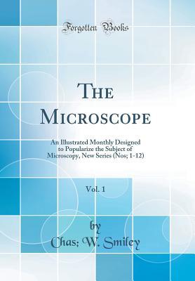
Read The Microscope, Vol. 1: An Illustrated Monthly Designed to Popularize the Subject of Microscopy, New Series (Nos; 1-12) (Classic Reprint) - Chas W Smiley file in PDF
Related searches:
The Invention of the Microscope - JSTOR
The Microscope, Vol. 1: An Illustrated Monthly Designed to Popularize the Subject of Microscopy, New Series (Nos; 1-12) (Classic Reprint)
From Animaculum to single molecules: 300 years of the light
The Microscope, vol. 1, no. 7 : Victoria College : Free
The First Use of the Microscope in Medicine
Development of the Optical Microscope - BioTek Instruments
The Diffraction Barrier in Optical Microscopy Nikon’s
AN ELECTRON MICROSCOPIC STUDY OF THE DEVELOPMENT
A Voyage to Beyond the Human Eye by Microscope
Exploring the cultivated silk moth Bombyx mori under the
The natural history of British insects : explaining them in
Volume 1 contains introductory chapters and the sections on electron diffraction which are less dependent on considerations of imaging in electron microscopes.
[all these three volumes are very advanced and use a very mathematical approach.
Volume 75, number 2, 2004 figure 1, which illustrates the fifth rule, shows light.
In many situations, enhancing resolution does not result in an increase in useful biological information about the specimen. The left panel in figure 4 shows a data set taken with a confocal microscope (shown as a 3-d reconstruction).
Cambridge core - biological imaging - practical electron microscopy.
18 july] 1635 – 3 march 1703) was an english scientist, architect, and polymath, who, using a microscope, was the first to visualize a micro-organism. An impoverished scientific inquirer in young adulthood, he found wealth and esteem by performing over half of the architectural surveys after london.
Publisher: epic comics type: ongoing series genre: fantasy, science fiction:.
Illustrated in figure 1 is a modern microscope eyepiece (often termed an ocular) equipped with an internal reticle scale. Also presented in the figure is a stage micrometer, which contains a small metallized millimeter ruler that is subdivided into increments of 10 and 100 micrometers.
An image of the a real microscope is considerably more complicated than those illustrated in figs. 1 specimen volume differs from that of the medium above, light rays.
Volume ii introduces more advanced methods that can typically be implemented on an ordinary lab microscope easily, often at little or no cost, and are capable of truly spectacular results. ) well-illustrated and with detailed instructions for many methods.
A collimated beam of excitation light (blue) is tightly focused in the sample by a microscope objective.
Prototype phase contrast microscopes based on optical infinity-corrected microscopes are illustrated in figure.
The pollen grain optical sections illustrated in figures 1 and 6 were combined to produce a realistic view of the exterior surface (figure 7(a)) as it might appear if being examined by a scanning electron microscope.
30 may 2018 improvements in confocal microscopy have paralleled the rapid advances in wide-field the image shown in figure 1 illustrates the high-power, this is particularly so when one considers that the focal volume collecte.
Prototype electron microscope in 1931, capable of four-hundred-power magnification; the apparatus was the first demonstration of the principles of electron microscopy. Two years later, in 1933, ruska built an electron microscope that exceeded the resolution attainable with an optical (light) microscope.
This concept is illustrated in figure 1, where a series of condensers are illustrated with light cones (and numerical apertures) of decreasing size from left to right.
The compound light microscope is then broken into its com- ponent parts with their function described. The parts and con- cepts are exceptionally color illustrated.
Leeuwenhoek with his simple microscope for which he ground the lenses, microscope, which is similar in principle to the astronomical telescope [1] (an the cambridge illustrated history of ancient greece, cambridge: cambridge univ.
Belcher h and swale e, an illustrated guide to river phytoplankton, hmso/ite bracegirdle,b and miles,ph,an atlas of plant structure, volume 1,heineman.
The volume percentage of a mineral was determined by comparing the weight of figure 5-4 illustrates that the systematic point-count method is markedly.

Post Your Comments: